

AI video microscopy delivers real-time, high-resolution imaging of living cells

Time to nerd out a little bit. Researchers at the University of California San Diego are using AI to peer into the inner lives of cells. The system, which extrapolates movement and images based on real data, “captures images that are twice as sharp as conventional microscopes and is fast enough to play as smooth video.”
The paper, which appeared in the Nature Communications, describes a process that “looks” at a real image of a cell or other biological matter and creates high-quality images without “false details.”
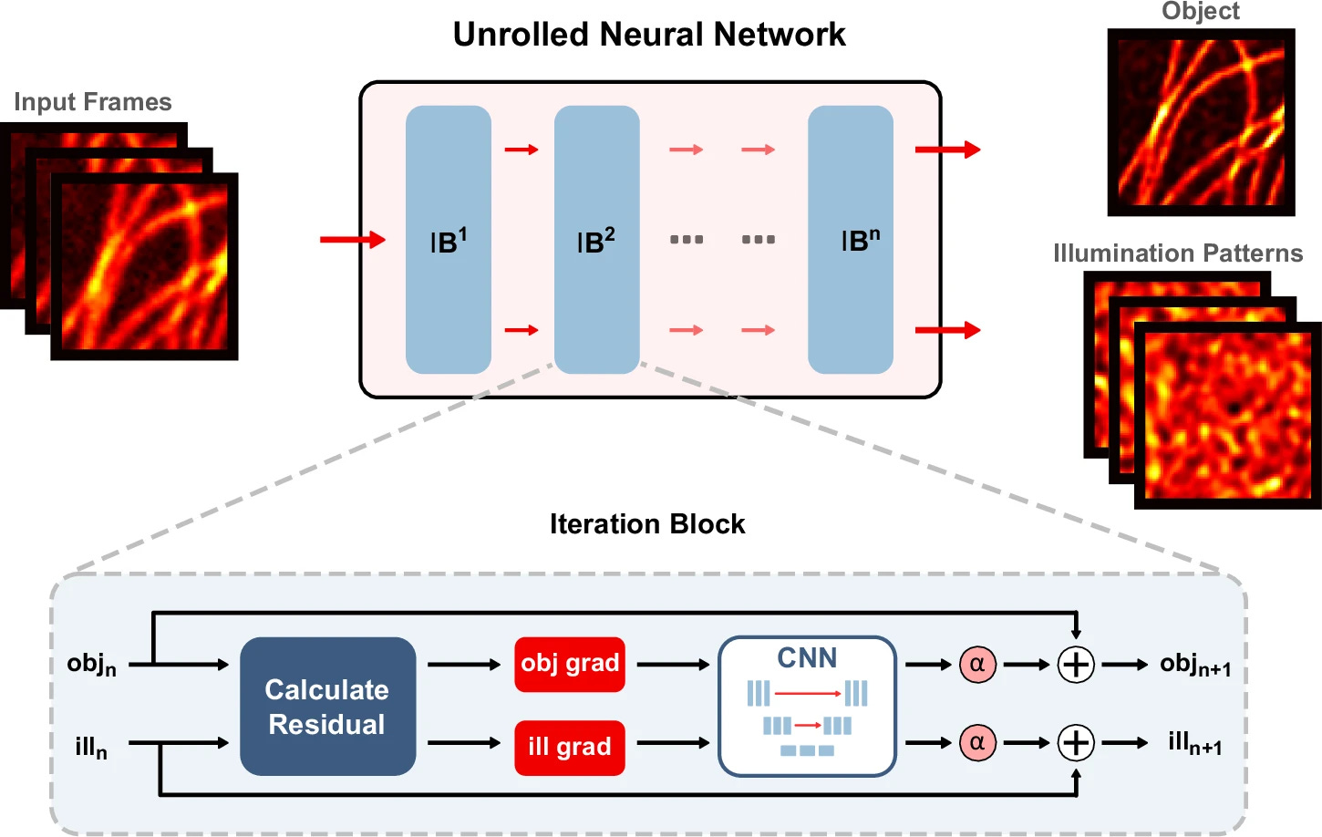
This work builds on structured illumination microscopy, or SIM, a method people have used for years to get sharper images out of living cells. The idea is simple: you shine patterned light on a sample, take a handful of images, and combine them to pull out detail you would not otherwise see. It works well because it is fast and does not blast the sample with too much light, which matters when you are looking at cells that are still alive or are sensitive to UV.
Traditional SIM systems need very tight control over the light patterns. If the pattern is even slightly off, the image quality drops. On the other side, the simpler versions that use random patterns avoid that hardware headache but pay for it later in speed.
The group led by Zhaowei Liu built what they call unrolled blind-SIM, or UBSIM. They rewired the reconstruction step so the system learns how to interpret the incoming data while staying tied to the underlying physics. The result is speed which results in image reconstruction happens hundreds or even thousands of times faster.
Then there is the problem of hallucinations. A lot of AI-based imaging tools look impressive but can invent structure that is not there. That is a problem if you are trying to study something as delicate as a cell. UBSIM avoids that trap by anchoring the model in how light actually behaves.
“One of the most exciting advancements with this algorithm is the removal of artifacts and hallucinations,” said study author Zachary Burns, an electrical and computer engineering Ph.D. student. “Currently, many neural network-based models can imagine fake structures when they are applied to new data. This is a major problem for scientists who use these AI models—they need to trust that the structures in the cell they are observing are real. By integrating optical physics, our model removes these issues and builds confidence that it can be used accurately.”
In practice, the system can produce high-resolution video of live cells at around 50 frames per second. That is fast enough to watch structures inside the cell change in real time, things like the endoplasmic reticulum moving and reshaping. It turns what used to be a slow, staged process into something closer to watching a live feed.
Liu’s point is that this changes how people use the tool. If the image shows up instantly and you do not need to babysit the system, it starts to feel like a normal microscope, just with much more detail. This lowers the quality barrier, speeds up experiments, and makes it easier to see something the moment it happens instead of after the fact.